

Plants consist of roots, stems, leaves, flowers, and fruits, which all work together to support their growth and produce food for consumption. Each part serves a specific function and is part of its identity as part of an organism’s ecosystem.
Demonstrate two leaves from various plant families and invite students to draw them, explaining that although they look different, they serve the same function.
Chloroplasts
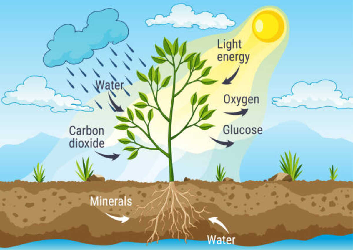
Chloroplasts are organelles found within plants that perform photosynthesis, which plants convert sunlight into food and energy for growth and survival. Commonly referred to as the “kitchen of the plant”, chloroplasts produce and store the necessary nutrition that keeps life forms alive and kicking.
Chloroplasts contain pigments such as chlorophyll that give plants their green hue, producing and storing chemical energy in the form of ATP (adenosine triphosphate). Chloroplasts can be found in all plant cells but are particularly abundant in leaf-producing cells, with approximately half a million chloroplasts per square millimeter of leaf tissue, which amounts to billions total in an entire plant!
Like mitochondria, chloroplasts may have arisen from oxygen-producing photosynthetic bacteria that were endocytosed into primitive eucaryotic cells and began producing oxygen. Like mitochondria, both utilize sunlight-driven ATP synthesis through light reactions taking place within their membranes to have energy for life processes; however, unlike mitochondria, the chloroplast genome only contains 120-200 kilobases of DNA, while their outer membrane allows freely permeable passage for small molecules and has transmembrane channels to import larger proteins encoded from nuclear DNA.
Inside the chloroplast are structures known as thylakoids. Each thylakoid is lined with cyanobacteria cells and enclosed by an extra-thick membrane with numerous folds; pigments like chlorophyll and carotenoid help absorb light energy for photosynthesis, and it is connected by stromal lamellae, which serve as bridges between them to keep them spread apart and maximize photosynthesis efficiency.
Thylakoids are connected to the chloroplast’s stroma by protein complexes that function in an electron transport chain. This chain converts light energy absorbed by thylakoids into chemical energy in the form of ADP and NADPH; their production fuels processes that make plants green like producing sugar from carbon dioxide and water as well as producing oxygen.
Stomata
Stomata are microscopic openings in the epidermis of leaves that allow for gaseous exchange, including carbon dioxide uptake and oxygen release, photosynthesis, and transpiration rate control. Stomata are activated by temperature, humidity, hormone signals, CO2 concentration, or other environmental triggers such as temperature or CO2. Opening and closing affect gas exchange rates directly and are thus critical elements in plant growth.
The stomatal opening occurs as a result of changes in internal pressure within guard cells that surround each pore, caused by an increase in water flowing into their vacuoles via osmosis and forcing their walls outward, expanding and pulling at inner wall borders to raise stoma opening. Once this water has left, guard cells contract back down, forcing its closure back onto each pore.
Depending on how their guard cells are enclosed, there are various types of stomata. Pilocytic stomata have two guard cells encircled by one subsidiary cell that contacts ordinary epidermis cells; diacritic stomata have two guard cells each encased by two lens-shaped subsidiary cells; while hemiparasitic stomata have two narrow guard cells surrounded by one wide cell similar to their guard cells but broader still.
Stomata are most prevalent among bryophytes in those liverworts, which resemble the leaves of flowering plants with their convoluted epidermis and porous interior. However, unlike genuine leaf stomata found elsewhere, those found on those liverworts do not open and close according to dynamic movement, as seen with higher plants’ leaves.
Stomata are essential in the life cycle of all plants as a primary means to absorb carbon dioxide and release oxygen in their surroundings. They regulate water uptake, adapting to changing weather conditions by opening or closing depending on which is necessary, regulating the uptake of water intake and adjusting as needed to changing weather conditions – thus making stomata an integral component in maintaining plant survival over the centuries.
Leaf Mesophyll Tissue
Leaf cells are uniquely tailored to capture and convert sunlight energy. Their thin walls and dense packing of chloroplasts, organelles that capture light energy for photosynthesis, make for efficient sunlight capture. Chlorophyll absorbs blue and red wavelengths of light which gives leaves their characteristic green hue. However, as daylight hours decrease during autumn, there becomes less solar energy available for photosynthesis; when this happens, chloroplasts degenerate, and their leaves change from green to yellow or orange in hue.
Mesophyll tissue comprises the area between the upper and lower epidermis layers, where mesophyll cells are organized into two distinct regions: palisade parenchyma is found beneath the upper epidermis layer with columnar cells oriented perpendicularly towards the leaf surface,. In contrast, in the lower part of leaf there exists irregularly-shaped cells called spongy parenchyma that collect light energy for capture by bundle sheath cells that contain mesophyll cells and have mesophyll cells also known as bundle sheath cells are highly specialized cells which specialize in light energy capture from light energy sources.
Palisade parenchyma cells are densely packed, long cells filled with chloroplasts that make up the top layer of mesophyll and are highly efficient at capturing light energy as their chloroplasts are situated directly in front of leaf surfaces allowing for increased exposure when sunlight moves across the sky. They have explicitly evolved for light capture because their chloroplasts can quickly move to intercept more sunlight as the sun moves across its path across the sky.
Spongy parenchyma cells have more irregular shapes and are packed with chloroplasts; however, their purpose is less focused on light energy capture than palisade cells, and they’re not as tightly packed together as palisade cells are. Mesophyll cells of leaves are lined by thick-walled vascular bundles consisting of xylem and phloem that transport water from roots to mesophyll cells for photosynthesis; conversely, phloem transport sugars produced during photosynthesis back to roots again for recycling by photosynthesis back through photosynthesis phloem.
Stomata are located within the vascular bundle. Their presence provides mechanical support and some types of photosynthesis. Furthermore, this structure is highly optimized for fluid transport – water and minerals move up through xylem, and sugar produced in leaves is transported out through the phloem.
Leaf Cross-section
Leaf cross-sections are cut through the leaves of plants to reveal their cellular structures, helping students gain insight into how plants operate by providing visual representations of cell parts and functions. Furthermore, cross-section diagrams can demonstrate how all components work together to produce food for the plant.
Leaves of vascular plants come in many different shapes and sizes. From the simple green blade of a pine tree to highly divided leaves on ferns, all leaves share one common trait: the blade or lamina which connects directly to the stem using a petiole; veins that conduct water and food around the leaf are known as vital zones.
Cross-section of lilac leaf showing midvein (large central vein), labeled with its water-conducting tissue xylem and food-conducting tissue (phloem). Stomata on the lower part are indicated with dots. The outer layer is called the epidermis; inside, the mesophyll comprises palisade cells and spongy parenchyma cells that make up photosynthetic tissues of mesophyll; Finally the veins themselves are protected with bundle sheaths encasement.
The left and right images above display plant adaptations for aquatic and xeric environments, respectively. Water lily leaves (Nymphaea) have cuticles to control water loss during photosynthesis; on Ficus trees (Figs), their stomata contain guard cells to prevent any entry or exit of moisture, respectively. Furthermore, both mesophyll cells have guard cells to remove unwanted moisture vapor. In both instances, their mesophyll is tailored specifically for their environments with numerous stomata and vascular tissues to increase the efficiency of photosynthesis.
Understanding the role of leaves in photosynthesis requires an in-depth knowledge of their layered structures. An excellent way to do this is with an online tool like Edraw Max Online that makes drawing more accessible, giving students a better grasp of each part of a leaf’s function.